
The cavernous sinus corresponds to a large vein located at the base of the skull. His mission ? Drain blood from facial veins. Associated pathologies, treatments… We take stock with Dr Caroline Apra, Head of Neurosurgery Clinic at Henri Mondor Hospital.
Definition and anatomy of the cavernous sinus
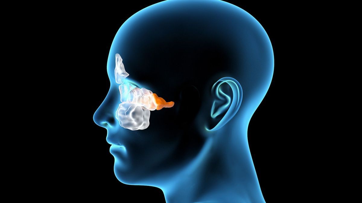
Where is the cavernous sinus located? What's going on inside? (Nerves, internal carotids…)
The cavernous sinuses are two large veins located on the anterior base of the skull, behind the eyes, on either side of the sphenoid bone. These veins drain blood from the veins in the brain. Inside the cavernous sinuses pass the internal carotid arteries which irrigate almost the entire brain, as well as the cranial nerves, in particular the oculomotor nerves which ensure the movement of the eyes and part of the trigeminal nerve which is responsible for facial sensitivity. The cavernous sinus has nothing to do with the other sinuses (frontal, maxillary, sphenoidal, ethmoidal), filled with air which open into the nasal cavities and become infected when you have a cold.
Physiology of the cavernous sinus
The role of the cavernous sinuses is to drain blood from the veins of the brain.
Cavernous sinus pathologies
Thrombosis or thrombophlebitis of the cavernous sinus
Very rare, cavernous sinus thrombosis is characterized by the formation of a blood clot (thrombosis) in the cavernous sinus. This pathology is generally linked to spread of bacteria linked to an infection of the face, orbit or sinuses. It manifests itself with severe facial pain, visual disturbances and high fever. Diagnosis is based on symptoms and brain MRI. Treatment is based on the administration of antibiotics.
Cavernous sinus tumor (meningioma, etc.)
A benign tumor can nestle in the cavernous sinus. The most common is meningioma, which develops from the meninges, the membranes surrounding the central nervous system, brain and spinal cord. “If the meninges are diseased, they can gradually thicken and form a meningioma, more or less extensive, in the cavernous sinus”informs the neurosurgeon.
Most meningiomas are asymptomaticthey are discovered incidentally during a radiological examination (CT or MRI) carried out for another reason. “Depending on its location, the tumor can cause headaches, convulsions or epileptic attacks, altered eye movements, loss of vision, speech problems, change in behavior, dizziness or even balance problems”specifies the specialist.
Post-traumatic brain pathologies
“In the event of an impact at the base of the skull, fistulas or dissections can occur. Consequently, the vessels which pass inside, in particular the carotid, can be damaged and come into communication with the interior of the skull. Sinus damage can cause hemorrhage. congestion in the eye (red, swollen, throbbing eye), because the carotid artery beats directly into the eye”explains Dr Caroline Apra.
Scanner, MRI: examinations of the cavernous sinus
The reference examinations for exploring the cavernous sinus are CT scan, MRI (magnetic resonance imaging), and sometimes arteriography with or without injection of contrast product. In some cases, blood cultures may be necessary.
Treatments and prevention of cavernous sinus problems
Treatment depends on the cause. The management of cavernous sinus thrombosis is based on the administration of antibiotics, sometimes combined with corticosteroids and anticoagulants. Surgical drainage may also be necessary if the sphenoid sinus is affected.
In the event of a tumor, treatment is essentially based on radiotherapy. “It’s an area that is difficult to operate on because there are too many things passing through it, the risk of stroke is very high. At the same time, certain hormonal treatments are contraindicated,” points out the neurosurgeon.