
Histology is a discipline that involves studying biological tissues to help diagnose sick patients or to find new treatments in medical research. Who can perform a histological examination? How is a histological examination carried out? What is the difference between cytology and histology? Answers with Hervé Gegout, histologist at the Strasbourg Biomedicine Research Center.
Histology is a specialty that involves studying the microscopic structure of tissues and organs. “This can be tissues of skin, bones, teeth, organs, cartilage, etc. Everything that makes up the human body can be studied during a histological examination”, specifies Hervé Gegout. It is used in biological research and medicine.
What is the purpose of histology?
Histological examination can be used to clarify a diagnosis. For example, in oncology, histology makes it possible to determine whether a tumor is malignant or not, or to provide information on the characteristics of a malignant tumor (its type, its stage, its grade, its aggressiveness).
Histology is also used in biological research to develop new treatments for many diseases. “Recently, I worked on the development of a patch allowing cartilage regeneration. Clinical trials, i.e. studies carried out on human beings, should begin soon. This advance could make it possible to treat people suffering from osteoarthritis”, says the histologist. Still in research, histology represents one of the means of studying the mechanisms of tumor development.
How is a histological examination carried out?
The histological examination can be divided into several stages.
Sampling
The first step is to take very small tissue using a biopsy. “The patient must always give consent before undergoing a biopsy. He can refuse. This type of collection is highly regulated in Europe to avoid excesses.”, insists the histologist. For histological examination, the tissue can also come from surgical specimens (partial or complete organ resections).
Fixation
Once collected, the tissues must be quickly immersed in a fixative liquid. Fixation is essential to preserve the tissues in a state as close as possible to their living state (before collection). The most commonly used fixative liquids are formalin or Bouin’s liquid. The fixation duration varies depending on the volume of the samples.
The dehydration
Dehydration consists of removing the water contained in the cells of the removed tissues. This step is necessary to then be able to make a fine section of the tissue without losing its initial cellular structure (which could happen when the plasma membrane which contains liquid ruptures). To do this, the sample is passed through alcohol baths of increasing concentrations (first alcohol diluted to 50 degrees to go up to absolute alcohol at 100 degrees).
Impregnation
The sample is passed through a liquid containing xylene or toluene to eliminate traces of absolute alcohol.
L’inclusion
The sample is immersed in melted paraffin which infiltrates all the cells in the sample. This step, which lasts several hours, will allow the production of thin sections (with a thickness of 2 to 5 µm) subsequently.
Blocking
The paraffin is poured into a small metal mold to be cooled in a freezer overnight. We then obtain a hard paraffin block in which the rigid sample is located.
Carrying out histological sections
The paraffin block is passed through a device (microtome) which makes it possible to produce sections of 2 to 5 µm arranged in regular series in the form of a ribbon. The histological sections are made in three stages:
- The spread: the paraffin ribbon segments are placed on a glass slide containing a spreading liquid;
- Collage : the glass slides are placed on a hot plate for 15 min at a temperature of 40°C;
- Drying : the glass slides are tilted and dried using absorbent blotting paper.
Deparaffinization
This step consists of removing the paraffin present around the sample. To do this, the glass slides are placed on a heating plate (45 to 60°C) for 15 minutes to liquefy the paraffin.
Rehydration
Rehydration consists of eliminating intracellular paraffin, by immersing the slides in alcohol baths of decreasing degrees (from 100° alcohol to 50° alcohol), then in distilled water.
Coloring
The coloring makes it possible to differentiate the different elements which constitute the tissues to be analyzed (nucleus, plasma membrane and cytoplasm). “There are a lot of colors. The stain chosen by the histologist will depend on the type of tissue to be analyzed. The coloring will not be the same for a skin sample as for a bone sample”, explains Hervé Gegout.
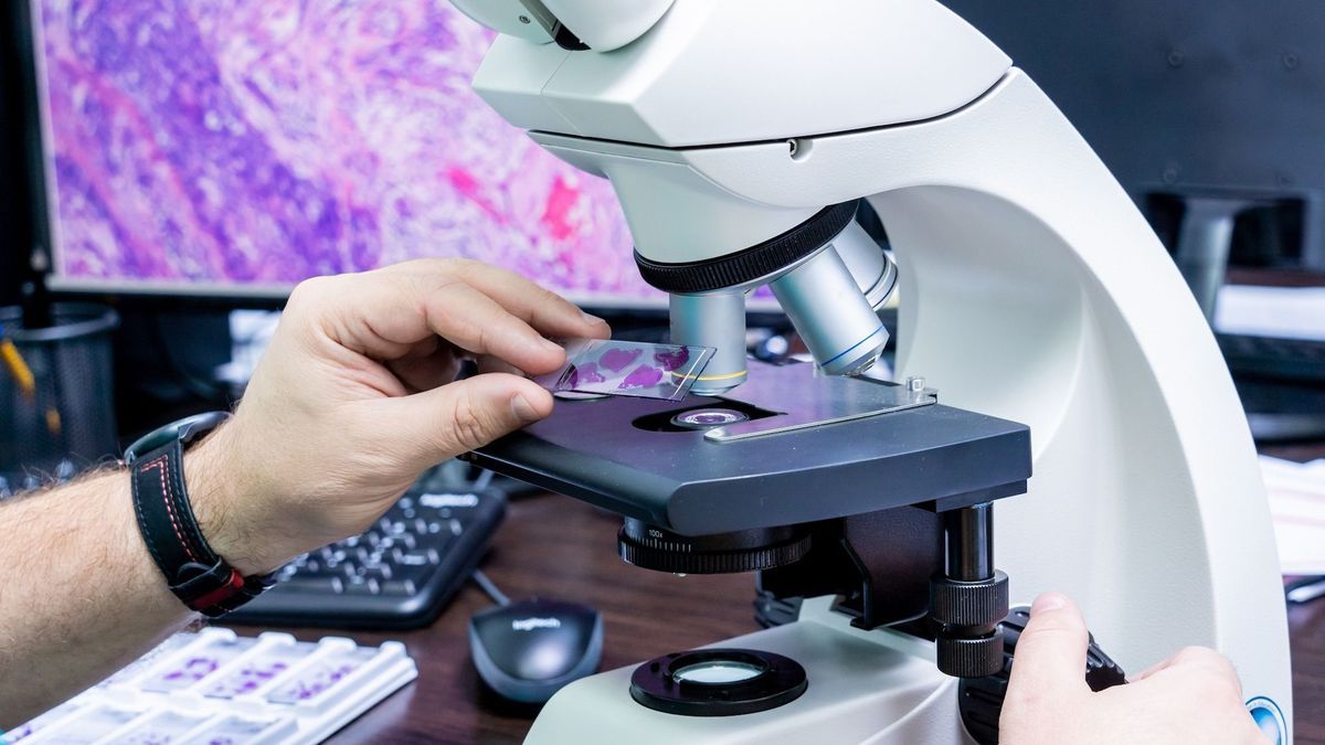
Assembly and observation under the microscope
Once colored, the biological tissue sections are mounted between slide and coverslip, which makes it possible to obtain a histological preparation (or “histological slide”) ready to be observed under an optical microscope.
What is the difference between a histologist and a pathologist?
The histologist is generally responsible for preparing the histological section. Its role is therefore to carry out all the steps mentioned above, except for tissue collection. The anatomopathologist’s mission is to examine the histological section under a microscope to identify and analyze anomalies linked to a disease.
The histologist working in the research field may also test drugs on organoids, which are miniature, simplified versions of an organ, made in vitro in three dimensions and which have realistic microanatomy. “To create organoids, we take human cells, dissociate them, then put them in culture to reconstitute the desired cellular model. It could be a mini liver, a mini lung, a specific cancerous tumor. This makes it possible to observe how an organ functions and reacts to different treatments, in an in vitro culture medium.”, explains the histologist.
By imitating the structure and functions of organs, organoids open the way to numerous applications: testing drugs, personalizing care or even improving cell therapy.
What is the difference between cytology and histology?
Cytology is the study of the constituents of the cell while histology studies biological tissues (association of cells of the same type).
Histology applied to oncology
In oncology, histology makes it possible to define the appearance of cancer cells which are compared to healthy cells. The histological examination of cancer cells will thus define the grade of the cancer which corresponds to the speed at which the cancer will progress and the risk that it will spread to other parts of the body. Defining the grade of a cancer can help predict the effectiveness of treatment and the outcome of the disease (prognosis). For some types of cancer, the grade is used to establish the stage. The stage of a cancer is its extent, that is, the amount of cancer cells present in the body and their location when initially diagnosed.