
Due to its size and complexity, the brain tends to obscure its partners within the encephalon. Among these, the midbrain. Located at the base of the brain, this nerve center supervises many vital functions and its name is associated with certain degenerative diseases, such as Parkinson’s disease.
What is the midbrain?
The midbrain, or midbrain, is located in the cranium. It corresponds to the upper part of the brain stem which is made up of two other structures: a protuberance, called the bridge, placed under the midbrain, and the myelencephalon, or medulla oblongata.
Above the midbrain, we recognize the diencephalon and especially the brain which appears to be placed on the midbrain – like Atlas carrying the world. Except that the midbrain has a trapezoid shape.
The midbrain itself is made up of three main structures:
- The cerebral peduncles which form two swellings;
- The tegmentum, seat of substantia nigra pars compacta (or locus niger), or the substantia nigra, of a certain number of cerebral nuclei and cranial nerves and of part of the reticular formation;
- The tectum, the upper part, as its name (“roof”) suggests.
Its structure is extremely complex, explains Professor David Devos, neuroscience researcher, neurologist and pharmacologist at the University of Lille, Lille University Hospital, Inserm, who compares the midbrain to a “carrefour” : “It is an area where major highways pass between the brain and the spinal cord. It houses numerous nuclei, cerebral and cranial nerves, which play very important vital and strategic roles..
The midbrain is also linked to the cerebellum which adjoins it. Exchanges take place via the tegmentum. It also forms the link between the cerebellum and the diencephalon.
The midbrain is vascularized by the two posterior cerebral arteries and a branch of the basilar artery, located at the base of the skull – we also speak of the truncus arteriosus.
What are the functions of the midbrain?
The midbrain – just like the diencephalon – is a phylogenetically very old structure that is found in all animals. It can be considered one of the most primitive centers of the central nervous system. As a result, Professor Devos recalls, “it is involved in the control of basic situations, related to major automated functions, such as walking, sleeping, breathing, etc.”.
In detail, the midbrain plays a major role in regulation:
- Automatic, reflex or stereotyped motor skills (speaking, laughing, walking, etc.);
- Postural stability and tone;
- Cardiovascular centers;
- Respiratory centers;
- Vigilance and sleep-wake cycles – the reticular formation, present along the entire length of the brain stem, is singularly involved in these vital functions, especially in its ascending component. Damage to the reticular formation can lead to coma;
- Pathways of exchange with the cerebellum, which Professor Devos likens to “a construction site manager”particularly with regard to locomotion and muscle tone;
- Visual pathways, and in particular oculomotor skills.
“The midbrain is a large place of integration of information which rises from the spinal cord (deep sensitivity) and descends from the brain (motor skills)”summarizes the researcher.
What are the pathologies associated with the midbrain?
The midbrain is, essentially, associated with two major degenerative pathologies, notes Professor Devos: Parkinson’s disease and progressive supranuclear palsy (PSP), which is a Parkinsonian syndrome. The PSP is reminiscent of “Parkinson”, but the similarity ends there. “It is very severe, with a life expectancy limited to 5-7 years.describes the specialist.
- Parkinson’s disease : this neurodegenerative disease which is accompanied by motor skills problems (slowness, stiffness, reduced range of movements, etc.), difficulty in writing, grasping objects and concentrating, or even tremors is explained by a decreased production of dopamine, a neurotransmitter secreted by the substantia nigra. “The affected area degenerates, leading to Parkinson’s disease”, declares the neurologist. The pedunculopontine nucleus, also located in the tegmentum, also plays a role in Parkinson’s disease. It is filled with acetylcholine, a neurotransmitter involved in muscle activity, balance, sleep and even memory. In the event of a deficiency, all of these functions are affected.
- Progressive supranuclear palsy: PSP is a rare disease. “The first symptoms appear around age 70, on average, explains Professor Devos. One of the characteristic signs is the inability for the patient to raise or lower their eyes, like vertical paralysis. Among the other manifestations: progressive walking disorders with extremely traumatic early falls, speech and swallowing difficulties and general slowing down, which can also affect the person’s intellectual abilities. “On imaging, the midbrain appears atrophied, in cases of PSP.adds our interlocutor.
In addition to these two neurodegenerative diseases, the midbrain can be affected by:
- Brain stem tumors, originating in or impacting the midbrain. These tumors can be primary or secondary, and occur in a malignant or benign form;
- A cerebrovascular accident (CVA)capable of impacting certain of its functions, such as eye disorders or sensitivity, permanent drowsiness or motor after-effects, illustrates Professor Devos;
- A serious viral or bacterial infection (herpes, measles, chickenpox-zoster), causing rhombencephalitis (inflammation of this part of the brain). The origin can also be immunological, autoimmune or post-infectious (hepatitis A or B, for example).
What are the diagnostic tests?
According to Professor David Devos, there are several ways to examine the midbrain, “direct, indirect and functional”.
Regarding imaging exams:
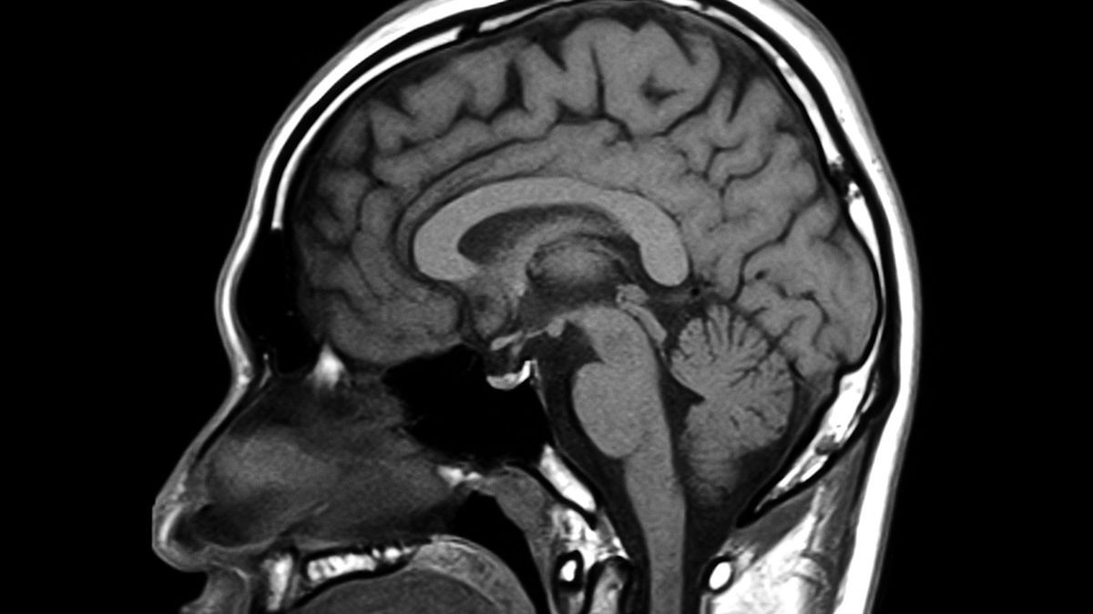
- MRI for the direct way. “The higher the field of MRI, 3 Tesla, and soon 7, the more precisely we see the structures of the midbrain. The approach is almost anatomical.notes the neuroscience researcher.
- Functional nuclear imaging which aims to see if the midbrain activates normally or not. Two devices with injection of radioactive glucose as a tracer: the PET-scan or the TEP-scan, for position emission tomography. They are used in the detection of dementia and cancers, in particular.
Two indirect approaches: the electroencephalogram which focuses on cortical activity and the recording of evoked potentials to study nerve conductions, motor pathway and sensory pathway. In the same vein, let us also mention transcranial magnetic stimulation.
The clinical examination also counts, obviously. A questioning is enough to make the diagnosis of Parkinson’s disease. Likewise, adds the neurologist, “the inability to look vertically indicates progressive supranuclear palsy in a senior. The same goes for suspected encephalitis – which should lead to the emergency room. Serious neurological symptoms include confusion, difficulty alertness, and even problems walking and vision – in addition to fever with headache. The clinical examination can be completed, among other things, by a blood test and a lumbar puncture.
What treatments for damage to the midbrain?
They will depend on the cause.
- Dopaminergic drugs for Parkinson’s diseaseto compensate for the loss of neurons.
- However, there is no treatment whose effectiveness has been validated. against progressive supranuclear palsy. But research is underway. “In neurodegenerative diseases, the idea today is to develop neuroprotective treatments, which currently only exist in Charcot disease. specifies Professor Devos;
- Chemotherapy and radiotherapy for malignant tumorsand according to the stage of the disease;
- Thrombolysis in case of strokefollowed by drug treatments and possible rehabilitation;
- Treatments for encephalitis will depend on its etiology. Reminder: to prevent certain viral or bacterial infections, consider vaccination.