
Spinal MRI or MRI of the spinal cord is an imaging test that allows you to explore the entire length of the spinal cord and spine. How does this exam take place? For what indications is it prescribed? Are there any contraindications? Here is everything you need to know about spinal cord MRI with Professor Jean Chazal, neurosurgeon.
What is a spinal cord MRI?
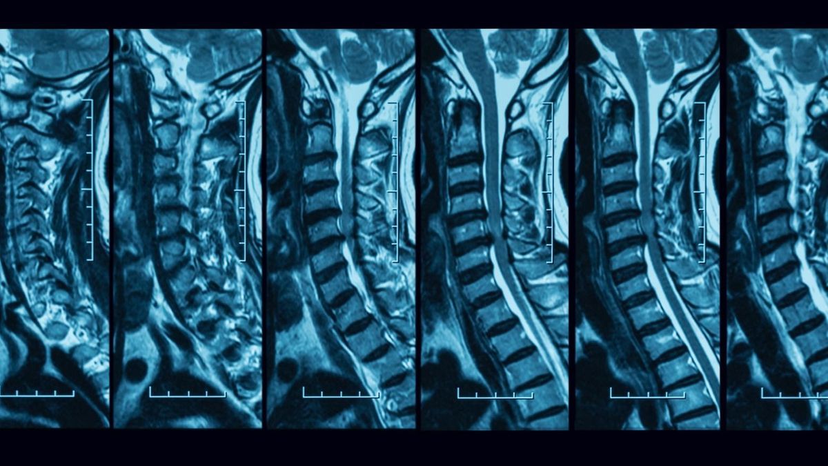
MRI is a medical imaging test which aims to explore the entire length of the spinal cord and spine. It is used to confirm or rule out injury to the spine and/or spinal cord.
As a reminder, MRI stands for magnetic resonance imaging. This is a radiology exam that uses a device that emits electromagnetic waves using a large magnet. When they are subjected to these waves, the hydrogen atoms making up the body’s tissues begin to vibrate. They then emit signals captured by a specific camera and transcribed into images on a computer screen. These images allow you to observe the interior of the human body, in 2 or 3 dimensions. MRI is particularly useful for visualizing the body’s soft tissues, namely the brain, spinal cord, viscera, muscles, etc. MRI also allows you to explore bones and joints.
In most cases, performing an MRI of the spinal cord requires the injection of a contrast product based on gadolinium, a metal. This product allows better visibility of body elements on images taken during the examination. “What is interesting with spinal MRI is that it allows us to distinguish the container, which is the spinal column, from the contents, which is the spinal cord. What no other exam allows you to do”indicates Professor Jean Chazal, neurosurgeon.
Why do a spinal cord MRI?
A spinal cord MRI is always prescribed after a careful clinical examination of the patient. It is indicated in cases of suspected damage to the spinal cord and/or spine. Damage to the spinal cord and spine can be linked to:
- A spinal cord tumor. Spinal cord tumors can be located in or around the spinal cord. “These tumors are usually benign but must be removed during surgery because they compress the spinal cord. Among them, we can cite astrocytoma, ependymoma or even neuroma.”, explains Professor Chazal;
- A vertebral angioma which is a vascular congenital malformation that develops over the years. It is a mass composed of blood vessels and fat that sits within the body of a vertebra. The vast majority of vertebral angiomas are asymptomatic but sometimes they can compress the spinal cord and cause significant pain;
- Syringomyelia which is characterized by the abnormal formation of one or more cavities inside the spinal cord. This is a rare congenital malformation;
- Trauma such as a spinal fracture. “In this case, spinal MRI will make it possible to assess the severity of the consequences of the fracture. Hemorrhage being the most serious consequence”, says the neurosurgeon;
- A spinal meningioma which is an uncommon tumor, most often benign;
- Metastatic epiduritis which results in compression of the spinal cord caused by the presence of metastases. It is a common neurological complication of cancer;
- Osteoarthritis of the spine characterized by chronic wear of the lumbar vertebrae. “This leads to the formation of bony beaks which compress the spinal cord.”, specifies Professor Chazal;
- Lumbar discarthrosis which is a common condition of the articular discs present in the back;
- An inflammatory pathology which can be multiple sclerosis or acute transverse myelitis.
Spinal MRI can not only help diagnose certain diseases but also observe their evolution. In fact, it can provide additional information to that obtained by an x-ray, an ultrasound or a scanner.
How is a spinal cord MRI performed?
Spinal MRI is an examination that lasts 20 to 30 minutes on average. If a gadolinium injection is necessary, it must be carried out a few minutes before the examination.
The patient lies on the machine table before entering a tunnel to perform the imaging examination. During the examination, the radiologist takes different images. He can dictate instructions to the patient if necessary. “For example, he can ask him to hold his breath for a few seconds to obtain more precise images”, explains the neurosurgeon. For their part, the patient can ask the radiologist to stop the examination if they do not feel well. As MRI is a noisy examination, noise-canceling headphones may be offered to the patient.
The radiologist can deliver the results at the end of the examination if he is able to analyze the images immediately or send them to the doctor prescribing the MRI who will in turn send them to the patient, if the interpretation of the results take more time. “This is done on a case by case basis. Generally, when there is nothing serious or even no lesion detected, the radiologist gives the results to the patient and comments on them at the end of the examination. In complicated or serious cases, the patient leaves without the results. He is informed that these will be communicated to him later by his doctor.”, indicates the specialist.
What are the contraindications for spinal cord MRI?
MRI is contraindicated for patients wearing metal equipment because the magnetic force produced during the examination is so powerful that it can damage or move these metal objects (pacemakers, implantable cardiac defibrillators, prostheses, transdermal patches, neurostimulators, surgical clips, metal shards in the eyes, etc.).
MRI is also contraindicated in pregnant women during the first trimester of pregnancy in order to limit, as a preventative measure, the potential risks for the embryo linked to the magnetic field.
For claustrophobic people, precautions can be taken by the medical team (prescription of a tranquilizer) to avoid discomfort.
If you have a tattoo on the area to be analyzed, namely along the spine, specify this when making your appointment. Tattooed skin may be slightly burned during an MRI. It is possible to prevent this type of incident by covering the tattoo with strips or an ice pack.
What are the side effects of spinal cord MRI?
Adverse effects of spinal cord MRI remain exceptional. This is a painless test with rare side effects. Gadolinium injection can cause reactions. In the vast majority of cases, they are temporary and benign:
- Feeling of warmth throughout the body or a “metallic” taste in the mouth.
- Nausea lasting a few seconds;
- Appearance of a small hematoma at the site of the bite;
- Subcutaneous leakage of contrast medium.
Some people may have an allergic reaction to the contrast agent requiring treatment.
Spinal MRI in multiple sclerosis
MRI is the reference imaging test for the diagnosis and monitoring of patients with multiple sclerosis. It will help to make the diagnosis at the beginning of the disease after the first clinical attack (manifestation of new symptoms for more than 24 hours, in the absence of fever) by highlighting the dissemination of plaques* in different locations of the brain (cerebral MRI). ) and sometimes the spinal cord. The MRI can also show that plaques have occurred at different times, helping to narrow down the diagnosis.
MRI is also used to monitor people with multiple sclerosis. It makes it possible to verify the effectiveness of a treatment in addition to the clinical examination by the neurologist. The latter can thus compare the latest MRI with the previous ones to see if the disease is progressing, if new plaques have appeared or if the disease is well controlled (perfect stability of already existing lesions).
*Lesions in which the protective sheath of neurons, myelin, is destroyed leading to degeneration of nerve cells.