
Rare but possible this genetic anomaly can be spotted during pregnancy. What is Down syndrome, can it be hereditary and how to detect it?
Definition of Down syndrome
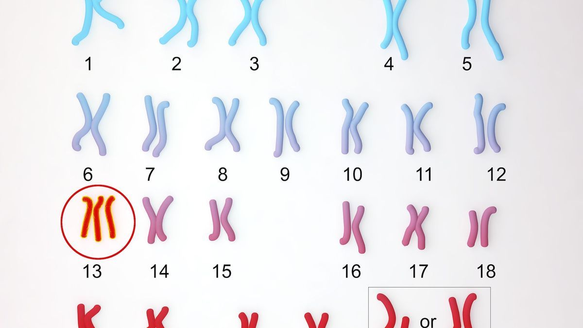
Trisomy 13 is a genetic anomaly corresponding to the presence of 3 chromosomes 13 instead of two. Which makes a total of 47 chromosomes instead of 46.
There are two types of trisomy 13:
Free trisomy 13
The three chromosomes 13 are distinct. Free trisomy 13 represents 80% of cases. This is the form of trisomy of accidental origin, linked to high maternal age.
Trisomy 13 by translocation
The third chromosome 13 is attached to another chromosome: another chromosome 13, or a chromosome 14, 15, 21 or 22. We speak of translocation robertsonienne. “In this case there is a greater risk of recurrence because one of the two parents may carry a translocation involving chromosome 13,” tells us Doctor Nicolas Gruchy, head of the genetics department at Caen University Hospital. “When there is an accidental free trisomy we say that the risk of recurrence is 1%. If it is a translocation trisomy, the risk is between 2 and 10%”.
What causes Down syndrome?
This anomaly most often occurs accidentally. Trisomy 13 is also linked to maternal age. “The risk of a fetus suffering from trisomy 13 will be greater in pregnant women aged 40 than in pregnant women aged 20. confirms the doctor. “We know that meiosis, the particular cell division which leads to the formation of gametes, particularly eggs, will be less efficient at the age of 40 than at the age of 20.”
No behavior or lifestyle habits of the pregnant woman could be linked to the occurrence of Down syndrome.
Signs, symptoms: how to recognize Down syndrome?
Free trisomy 13 and translocation trisomy will give the same severe clinical signs:
- Trisomy 13 is a polymalformative syndrome first described in 1960 by Klaus Patau, American scientist and geneticist. Trisomy 13 causes major malformations with facial dysmorphia (facial deformation), a small skull, a receding forehead, ulceration of the scalp, very small eyes, microphtalmiea wide, flat nose, a cleft lip and nostril (hare lip), a cleft palate;
- There are other malformations not visible on clinical examination, notably a holoprosencephaly which designates the absence of separation of the cerebral hemispheres. In cases of trisomy 13, there are very frequently heart malformations, limb anomalies including the presence of six fingers (hexadactylie), foot malformations (club feet), spinal malformations.
What is the difference with trisomies 18 and 21?
According to Santé Publique Europe, we can distinguish the two forms:
- The Down syndrome (Edwards syndrome) has an extra chromosome 18. It is characterized by growth retardation, visceral malformations affecting all organs including the heart in more than 90%, the limbs (club feet, hands and fingers folded and fixed), the neural tube (anencephaly, spina bifida) the tube digestive, kidneys, face (cleft lip and palate). More than 95% of affected fetuses die in utero;
- Down syndrome has an extra chromosome 21 (complete trisomy 21) or a supernumerary fragment of chromosome 21 (partial trisomy). It is characterized by mild to moderate intellectual disability, frequent muscular hypotonia and joint laxity. Congenital malformations are frequently associated (cardiac and digestive malformations in particular) as well as sensory disorders, epilepsy, leukemia, autoimmune and endocrine pathologies or premature aging, both sensory and neuro-cognitive (Alzheimer’s disease).
Trisomy 13: life expectancy and miscarriage
In utero fetal deaths are common in cases of Trisomy 13. In utero fetal deaths often occur in the first trimester of pregnancy. The malformations are more severe than others which means that some will not be compatible with pregnancy. “Nature is somehow well done in the face of severe malformations, recognizes the geneticist. The fetus evacuates through the natural channels, this results in a miscarriage. If the pregnancy is carried to term – which is rare today because we have prenatal diagnosis – life expectancy rarely exceeds one week. There are perhaps a few cases who will live a year or more but they will have very severe neurological manifestations.”
Public Health Europe suggests that “more than 95% of affected fetuses die in utero. Half of children die in the first month and 90% before 1 year from cardiac, renal or neurological complications.“.
How do you know if you have Down syndrome?
There are two circumstances for diagnosing Down syndrome:
1 / The malformation seen on ultrasound
Pregnancy ultrasound scans help to screen for trisomy 13. If malformations suggestive of trisomy 13 have been suspected during the ultrasound scan of the first or second trimester of pregnancy, the pregnant woman is offered, depending on the term of the pregnancy, two examinations allowing chromosomal analysis and diagnosis of trisomy 13:
- Trophoblast biopsy
The trophoblast biopsy is a sample that can be taken in the first trimester of pregnancy (from 10 weeks of pregnancy). The biopsy consists of taking a fragment of trophoblast (tissue which will subsequently become the placenta). “This trophoblast biopsy is done abdominally (like an amniocentesis), or vaginally, after placement of a speculum, indicates the specialist. This is an invasive sample with a risk of miscarriage (around 1%). Trophoblast analysis reflects the fetal karyotype. A rapid result can be available in 48-72 hours.
- Amniocentesis
This invasive examination is offered from 14 weeks of pregnancy. “If trisomy 13 is detected beyond 3 months, gynecologists will detect growth retardation or a cardiac or cerebral anomaly, which will prompt amniocentesis.” says the doctor.
Amniocentesis is a collection of amniotic fluid; the examination is carried out in a sterile manner using ultrasound guidance. The gynecologist will use a needle to pass through the abdominal wall to collect approximately 20 ml of amniotic fluid. The results are analyzed in the laboratory. It takes 3 weeks to receive the final results. “We have exams that allow us to respond to Down syndrome in 48 hours, would like to clarify, Doctor Nicolas Gruchy. What takes time is the cell culture, to obtain a complete karyotype.”
Amniocentesis does not require any special preparation for the pregnant woman before the examination. However, she is kept under observation in the office or hospital department for a few hours to check that there are no adverse effects post-intervention.
“Amniocentesis and biopsy allow the same chromosomal analysis to be carried out, indicates the specialist. The advantage of the biopsy is that it is earlier since it can be carried out between 10 and 12 weeks of pregnancy. Amniocentesis is a better reflection of fetal karyotype (analysis of cells from several tissues), it is not an appendix like the placenta. The risk of miscarriage is a little lower (around 0.5%).
No genetic analysis is carried out on first miscarriages.
2 / One of the two parents carries a translocation involving chromosome 13
In cases of trisomy 13 by translocation, when it is known that one of the two parents carries a translocation involving a chromosome 13 (after having carried out their karyotype), doctors can offer, from 10 weeks of pregnancy, a maternal blood sample. This non-invasive prenatal screening (NIPT) analysis of fetal DNA that circulates in maternal blood. When the NIPT is suggestive of trisomy 13, the suspicion must be confirmed by amniocentesis. We do not carry out the karyotype of the parents in cases of free Trisomy 13, which is more accidental.
Trisomy 13: pregnancy or abortion? How to do ?
In both cases, if trisomy 13 is confirmed, there is the possibility for the patient to request a Medical Termination of Pregnancy which is generally accepted by the multidisciplinary prenatal diagnosis center. “Doctors may accept termination of pregnancy for medical reasons in the event of a particularly serious and incurable condition, specifies Doctor Nicolas Gruchy. IMGs can take place until delivery.