
Muscle of the lower limb, located at the back of the leg, the soleus muscle constitutes, with the gastrocnemius muscle, the “triceps surae” muscle which allows the extension of the foot. Strong calf muscle, it can be affected by different muscular and tendon pathologies. Explanations with Dr Maurice Demol, general practitioner and sports doctor.
The soleus muscle and the gastrocnemius muscle together form the muscular mass located at the back of the leg, which is called “triceps surae” or more commonly “calf”. This set of muscles, essential for mobility, is heavily used and therefore at risk of injury.
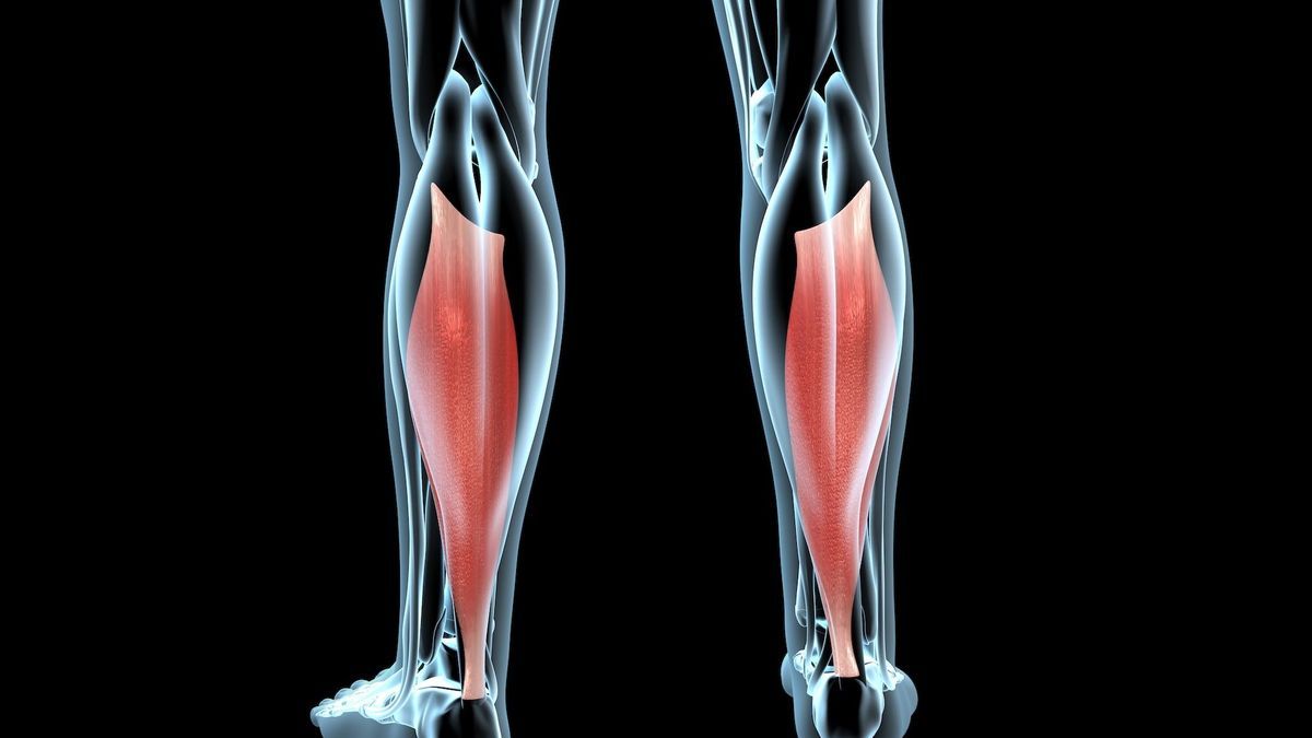
Anatomy, physiology: what is the soleus muscle?
The triceps surae is the posterior muscle group of the leg: it is what gives the calf its fleshy and chubby shape. It is one of the most powerful muscles in the body after the gluteus maximus and the quadriceps: we use it daily during our movements (walking, running, jumping, etc.). Extending from the femur to the calcaneus (i.e. from the thigh to the calf), the triceps surae is made up of two muscles:
- The soleus muscle : flat, thick and bulky muscle, located on the deep plane of the triceps surae, under the gastrocnemius, which connects the tibia and fibula to the calcaneus;
- The gastrocnemius muscle : superficial muscle, divided into two heads (medial and lateral, previously called the twin muscles).
Between these two muscles is the plantaris muscle, an inconstant accessory muscle, contributing little to the strength of the muscle group.
The triceps surae also plays a role in blood circulation since it promotes venous return. When walking, the repeated contractions of the gastrocnemius allow small volumes of blood to be ejected and empty the venous reservoir of the lower limbs.
Let us now look in more detail at the soleus muscle (from the Latin “solea” which means “sole of the foot”) through its characteristics
- Position : Bundle of the triceps surae muscle, the soleus muscle is contained in the superficial part of the posterior crural compartment, i.e. the posterior part of the leg, between the back of the knee and the ankle.
- Structure : This is a skeletal muscle, that is to say a muscle placed under the voluntary control of the central nervous system.
- Origin : The soleus muscle attaches to the posteromedial part of the fibular head, on the lateral edge and on the posterior surface of the fibula (also called fibula) by extending onto the interosseous membrane of the leg. It attaches at the level of the tibia on the line of the soleus muscle. These two insertion points are connected by the tendinous arch of the soleus muscle on which the muscle completes its attachment.
- Termination : It is inserted on the blade of the calcaneal tendon, or Achilles tendon, to attach to the tuberosity of the calcaneus.
- Innervation : Like the biceps femoris, its innervation comes from the tibial nerve.
The soleus muscle is a mono-articular muscle (meaning it only contributes to the movement of one joint). Located under the gastrocnemius, less visible, it is mainly composed of type I muscle fibers, more enduring and tonic. Unlike the gastrocnemius, the soleus muscle is therefore very resistant to fatigue. It has greater muscle activity when the knee is flexed.
What is his role ?
When we are standing, the soleus muscle is constantly active. “Maintaining balance in a standing position, it provides a power function (medial portion) and a stabilizing role for the ankle (lateral portion). Its main function, however, is to act as an extensor of the foot: it indeed allows ‘extend the foot bone and ensure plantar flexion’describes Dr Maurice Demol, general practitioner and sports doctor in Brussels.
Together, the gastrocnemius muscles and the soleus muscle are the main plantar flexors of the ankle (thanks to them we can point our foot). Together they contribute over 70% of the total strength of this movement. The other muscles, secondary flexors (posterior tibialis, toe flexors, etc.), only provide 30% of the plantar flexion force.
The soleus muscle is therefore the muscle for walking and jumping: it brings the back of the foot upwards, lowers the toes or raises the heel when the foot is on the ground.
According to a study published by researchers at the University of Houston in the United States on the iScience website, it finally helps to improve metabolic health: contracting this muscle, even in a seated position, allows you to burn fat. and regulate blood sugar levels.
What pathologies can affect the soleus muscle?
Pain relating to the soleus muscle is of muscular and tendon origin. However, the soleus muscle is more rarely affected than the gastrocnemius. “It is in fact less at risk of injury because it only crosses one joint (the ankle) and is only made up of one type of slow fibers. When an injury to the soleus occurs, it is generally less significant than for the gastrocnemius.”adds the specialist.
Here are the main pathologies and painful injuries that can affect it:
Muscle pain without damage
- Cramps: they correspond to an involuntary, painful and temporary contraction at the level of the soleus muscle;
- Contractures: However, these cramp-like pains are permanent. They appear during exercise or a few hours later. The calf is hard, can sometimes be swollen or have a bump. The pain increases when touched.
The duration of the pain varies: approximately a few minutes for cramps and rather 5 to 10 days in the case of contractures.
Muscle damage
- Elongation (or sprain) of the soleus: first stage of muscle injury, elongation is an elongation of muscle fibers beyond their elasticity, which creates micro-lesions (stretching of the muscle caused by micro-tears and leading to muscular disorganization). It causes deep pain or tension near the Achilles tendon. Symptoms tend to develop over time and initially manifest as calf fatigue, then gradually increasing pain.
- Tearing or breakdown: second stage of muscle injury, breakdown also corresponds to an elongation of the muscle fibers beyond their elasticity but this time leading to a rupture of these muscle fibers, linked to a greater mechanical stress than for elongation; leading to bleeding within the muscle, which can form a hematoma and edema. The incident was brutal with intense pain.
- Rupture or disintegration: last stage of muscle damage, it corresponds to a total rupture of the muscle (most often, following a sudden fall). The pain is very significant.
The “Tennis leg”
Tennis Leg is a common and potentially severe pathology involving the soleus muscle. It affects tennis players (hence its name) more frequently from the age of forty. A sudden start on a poorly warmed muscle is often the cause. “The medial gastrocnemius, which is bi-articular, will contract more strongly than the soleus (mono-articular), which will cause its aponeurosis to separate from that of the soleus., explains the sports doctor. This causes sudden pain and bleeding which can form a hematoma. “The hematoma, if it remains in place, risks becoming encysted and preventing the wound from healing properly. This results in long-term calf pain and weakness.”
Les tendinopathies
The Achilles tendon is formed by the union of the three parts of the triceps surae and can be the site of tendinopathy. This concerns all the pathologies which occur in the tendons and which manifest, initially (at the level of the soleus muscle and the Achilles tendon), by pain in the leg during exercise. . “Subsequently, when tendinopathy sets in, pain may be present even at rest.continues Dr Maurice Demol. Tendon stiffness sets in and a thickening of the tendon itself may be observed, making it more at risk of tearing. The origin of tendinopathy is varied. It can be linked to a repeated bad gesture, too heavy a sporting load, poorly adapted shoes, too little sports rest or poor hydration. It can also be a genetic predisposition, such as repeated poor posture during sports, particularly when running. Running is thus one of the sports most frequently causing Achilles tendinopathy.
Achilles tendon rupture
The Achilles tendon inserts directly into the calcaneus, the bone that forms the heel and is primarily responsible for flexion of the ankle. When there is a sudden tearing of the tissues in this area, we speak of “rupture of the Achilles tendon”. The signs are very typical, with sudden sharp pain and inability to walk.
What tests are necessary to make a diagnosis in case of pain in the soleus muscle?
Firstly, the clinical examination makes it possible to observe and evaluate the symptoms reported by the patient.
“A combination of palpations, muscle testing and stretching tests (testing pain, flexibility with passive ankle movements and stretching), carried out by a doctor (ideally a sports doctor) makes it possible to differentiate lesions of the gastrocnemius and soleus,…