
More commonly called dental panoramic radio, the orthopantomogram is an essential tool for the dentist before carrying out surgery or to check certain anatomical elements of the mouth. We take a look at this examination with Doctor Christophe Lequart, dental surgeon, national spokesperson for the French Union for Oral Health (UFSBD).
Orthopantomogram, definition
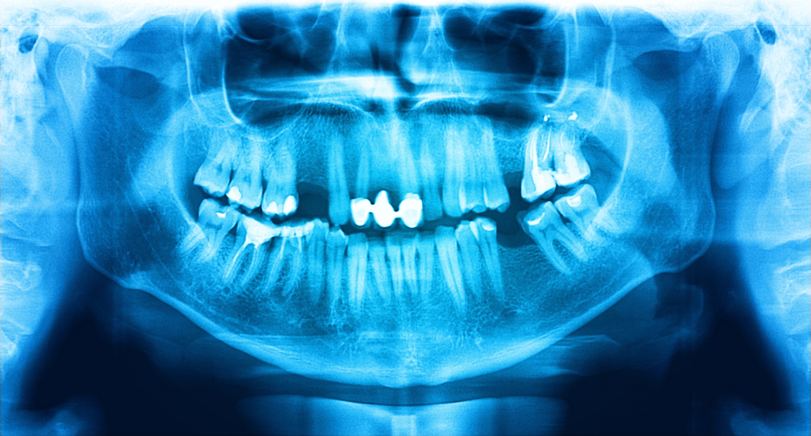
An orthopantomogram corresponds to a panoramic x-ray also called Panorex. The aim of this examination is to take an x-ray of the entire bony structures of the lower jaw and upper jaw for an overview, looking for an anomaly, trauma or tumor. . “The orthopantomogram makes it possible to inspect various anatomical elements at the bony level such as the mandible, the upper jaw bone, all the teeth and all of their roots. indicates Doctor Christophe Lequart, dental surgeon, national spokesperson for the UFSBD.
“We will also be able to look at the temporomandibular joints (the TMJ) which are the joints of the lower jaw. We will also have a vision of the right and left maxillary sinuses. The orbital cavities are also visible” explains the expert.
Why have a panoramic dental x-ray?
There are a large number of indications:
Tooth decay
The advantage of the orhopantomogram is to have an overview allowing an initial diagnosis to be made to identify carious lesions (in particular the beginning of caries) present on the teeth.
Periodontal diseases
The orhopantomogram also allows the practitioner or radiologist to make an initial identification of dental loosening problems and therefore periodontal diseases since it is possible to distinguish the areas where bone loss has taken place along the roots of the different teeth.
Cysts, granulomas or granulocysts
They are also visible on the orhopantomogram. “A cyst is hollow while a granuloma at the root is solid, says the doctor. These are infectious lesions that can appear at the ends of the roots when there is an infection of the tooth. These lesions are not particularly painful.
Evolution des dents
Panoramic x-rays can be done by the practitioner or radiologist to follow the evolution of the teeth. “This is the case if we want to check that a child has all the germs of future permanent teeth. specifies the specialist. “In the same way, panoramic radiography can inform us about the positioning of wisdom teeth in young adults and thus know whether or not they will potentially have room to grow correctly”. To find out more, read our article “Wisdom teeth: to extract them or not?”.
Positioning of the dental nerve canal
It is also good to check, before an operation to extract molars or wisdom teeth, the positioning of the lower dental nerve canal. “This nerve is very important because it supplies blood to the entire lower jaw. explains Doctor Christophe Lequart. “An injury to the dental nerve will lead to paresthesia or even a temporary or even permanent disappearance of the sensitivity of half of the mandible and half of the lower lip on the side of the affected dental nerve.
Oral-sinus communication
The panoramic radio can also provide information on the possibility of a communication oral-sinusal (direct communication between the roots of the upper molars and the interior of the maxillary sinuses) after operation and extraction of a tooth.
Infection des sinus
The panoramic x-ray is also capable of identifying an infection in a sinus which could be linked to a dental infection.
Joint wear
At the level ofTMJ (lower jaw joint), the panoramic X-ray makes it possible to identify joint wear and dislocation of the meniscus of this temporomandibular joint. “These are usually patients who complain of hearing clicking and noises when they open and close their mouth, specifies the dental surgeon. They complain of having their jaw stuck from time to time.”
Bruxism
In cases of bruxism, panoramic radiography is useful to see potential wear at the temporomandibular joint.
Various trauma
The orthopantomogram is also very useful in cases of accidental trauma. This is the case for jaw fractures, for example.
How to recognize a dental infection on an x-ray?
In all x-rays (dental panoramic or others) the diagnosis will be made based on a difference in clarity in the image. “It is this variation in degree between white and black that will make us analyze the different anatomical structures. indicates Doctor Christophe Lequart. “Vacuity translates into black or dark on the x-ray. A sinus is empty, so we will have something dark on the x-ray. A cavity, a cyst, an abscess appear dark compared to the bone clearer”.
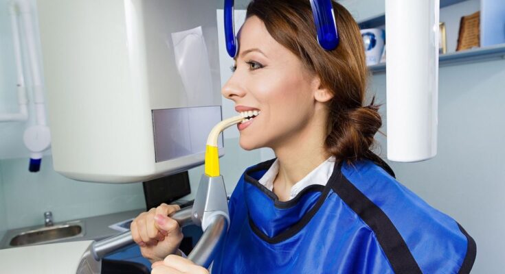
How is an orthopantomogram performed?
During the examination, the patient stands in an x-ray room (isolated booth) adjoining the dentist's office. The patient is positioned by the practitioner or his assistant in a particular way in order to obtain a good reading of the x-ray. The patient will then bite into a small wedge to have a space between the upper teeth and the lower teeth.
“This prevents the upper and lower teeth from overlapping when you close your mouth. indicates the expert. The x-ray camera will rotate for around twenty seconds around the patient at head level to perform the panoramic x-ray. During these few seconds, X-rays are projected like a conventional radio. The images taken by the x-ray camera arrive instantly on the dentist's computer screen.
What are the risks associated with radiation from an orthopantomogram?
The quantity of X-rays sent is much lower than before. “Radiology today has gradually moved from film to digital over the past 20 years. explains Doctor Christophe Lequart. “The advantage of digital sensors is that they send 10 times fewer x-rays than a film x-ray. Or 4 to 30 µSv for a panoramic, which corresponds to a few days of natural radiation or to a chest X-ray for the highest doses. Performing a panoramic x-ray on a pregnant woman does not involve any risk even if as a precaution she is placed with a protective apron and sometimes a lead collar to protect the thyroid.”
The dose of X-rays for each panoramic X-ray is adapted according to the age, size and morphology of the patient.
“An x-ray in a patient’s mouth is the equivalent of 4 hours of solar radiation,” would like to point out Doctor Christophe Lequart.
How much does a dental panoramic cost?
A panoramic X-ray at the sector 1 approved dentist costs 54 euros. Social security reimburses medical imaging procedures up to 70%. Be careful because the price may vary from one dentist to another. The sector 2 approved dental surgeon can in fact charge excess fees. Do not hesitate to make an appointment with your mutual to take stock of the conditions of your contract and the possible reimbursement of the remainder payable.
Where to do an orthopantomogram?
For your panoramic X-ray you can make an appointment with your dentist or at a dental radiology center.
Who can prescribe a panoramic dental X-ray?
Your GP may prescribe a panoramic x-ray. However, the dental surgeon can prescribe this X-ray and perform it himself.